Startseite

Datenschutz

Forschungstätigkeit


1. Discovery of structural elements of the nephrogenic zone and contained progenitor cell niches

Key features of the nephrogenic zone and contained progenitor cell niches in human fetal kidney are unknown. Consequently, actual research is focusing on spatial coordinates to recognize first signs of impairment of nephrogenesis in preterm and low birth weight babies.

Forschung2 The illustration shows in transmission electron microscopy mesenchyal (MES) and epithelial (E) cells within the renal stem/progenitor cell niche. Projections of mesenchyal cells line through the wide interface to contact epithelial cells.


Label of extracellular matrix within the renal stem/progenitor cell niche by innovative fixation


Interstitium
Unveiling of textural features
Conventional fixation of specimens by glutaraldehyde (GA) solution depicts an unobstrusive interstitial interface. In contrast, fixation of specimens by GA solution including cupromeronic blue, ruthenium red or tannic acid unveils earlier not visible structural details.

BlankPlus 
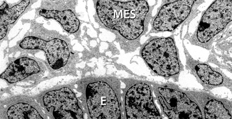
basal lamina with lamina rara (L.r.), densa (L.d.) and fibrioreticularis (L.f.).
BlankStern interstitial space.
Pfeil mesenchymal cell.
Spitze projection of mesenchymal cell.


W.W. Minuth (2023). Installation of the developing nephron in the fetal human kidney during advanced pregnancy. Molecular & Cellular Pediatrics 10:18.(PDF)

W.W. Minuth (2022). The interstitium at the developing nephron in the fetal kidney during advanced pregnancy - a microanatomical inventory. Molecular & Cellular Pediatrics 9(1):17(PDF)

W.W. Minuth (2021). The mutual patterning between the developing nephron and its covering tissues - valid reasons to rethink the search for traces left by impaired nephrogenesis. Mol Cell Pediatr 8(1):9.(PDF)

W.W. Minuth (2021). Nephronentwicklung im letzten Trimester der Schwangerschaft - prägend, klinisch relevant aber kaum erforscht. DER NEPHROLOGE 16:237-240.(PDF)

W.W. Minuth (2021). Microanatomy of the developing nephron in the fetal human kidney during late gestation. Annals of Anatomy. 236:151705. DOI:10.1016/j.aanat.2021.151705(PDF)

W.W. Minuth (2020). Shaping of the nephron - a complex, vulnerable and poorly explored backdrop for noxae impairing nephrogenesis in the fetal human kidney. Molecular & Cellular Pediatrics 7(1):2.(PDF)

W.W. Minuth (2019). In search of imprints left by the impairment of nephrogenesis. Cells Tissues Organs 207(2):69-82.(PDF)

W.W. Minuth (2019). Key features of the nephrogenic zone in the fetal human kidney – hardly known but relevant for the detection of first traces impairing nephrogenesis. Cell and Tissue Research 375(3):589-603.(PDF)

W.W. Minuth (2019). Structural and functional links between capsule and nephrogenic zone in fetal human kidney. J Pediatr Neonat Individ Med (JPNIM) 8(1):e080105.(PDF)

W.W. Minuth (2018). Reading first coordinates from the nephrogenic zone in human fetal kidney. Nephron 138(2):137-146.(PDF)

W.W. Minuth (2018). Action plan for prolongation of nephrogenesis in preterm and growth restricted babies: Explore ultrastructure of the nephrogenic zone, identify a molecular target, select a viable drug and find a path for administration. Drug Res 68(1):5-16.(PDF)

W.W. Minuth (2017). Concepts for a therapeutic prolongation of nephrogenesis in preterm and low birth weight babies must correspond to structural-functional properties in the nephrogenic zone. Mol Cell Pediatr 4(1):12.(PDF)

W.W.Minuth (2017). Reading first coordinates from the nephrogenic zone in human fetal kidney. Nephron doi: 1159/000481441.(PDF)

W.W. Minuth (2017). The rabbit nephrogenic zone in culture: past, present and future as a model to investigate causes of impaired nephrogenesis. J Pediatric Neonatal Individualized Medicine. JPNIM 6(1):e060111.(PDF)

W.W. Minuth (2017). View onto the nephrogenic zone before stem cell niches come apart: challenge for smart drug delivery. J Drug Res DEV 3(1):doi http://dx.doi.org/10.16966/2470-1009.127.(PDF)

W.W. Minuth, L. Denk (2016). Special morphological features at the interface of the renal stem/progenitor cell niche force to reinvestigate transport of morphogens during nephron induction. BioResearch Open Access 5(1):1-12.(PDF)

W.W. Minuth, L. Denk (2016). What is the functional background of filigree extracellular matrix and cell-cell connections at the interface of the renal stem/progenitor cell niche? J Pediatric Neanatal Individiualized Medicine 5(1):e50115.(PDF)

W.W. Minuth, L. Denk (2015). When morphogenetic proteins encounter special extracellular matrix and cell-cell junctions at the interface within the renal stem/progenitor cell niche. Anat Cell Biol 48(1):1-9.(PDF)

W.W. Minuth, L. Denk (2014). Separating and connecting properties of the interface within the renal stem/progenitor cell niche. Stem Cell Biol Research. 1:4.(PDF)

W.W. Minuth, L. Denk (2014). Advanced fixation for transmission electron microscopy unveils special extracellular matrix within the renal stem/progenitor cell niche. Methods Mol Biol. 1212:21-37.(PDF)

W.W. Minuth, L. Denk (2014). Structural links between the renal stem/progenitor cell niche and the organ capsule. Histochem Cell Biol 141(5):459-471.(PDF)

W.W. Minuth, L. Denk (2013). The interstitial interface within the renal stem/progenitor cell niche exhibits an unique microheterogeneous composition. Int J Mol Sci 14:13657-13669.(PDF)

W.W. Minuth, L. Denk (2012). Cell projections and extracellular matrix are crossing the interstttial interface within the renal stem/progenitor cell niche ? accidental, structural or functional cues? NEPHRON Exp Nephrology 122(3-4):131-140.(PDF)

W.W. Minuth, L. Denk (2012). Illustration of extensive extracellular matrix at the epithelial-mesenchymal interface within the renal stem/progenitor cell niche? BMC Clinical Pathology 12:16.(PDF)

W.W. Minuth, L. Denk, Ch. Miess, A. Glashauser (2011). Peculiarities of the interstitium in the renal stem/progenitor cell niche. Histochem Cell Biol 136:321-334.(PDF)

K. Schumacher, J. Klar, C. Wagner, W.W. Minuth (2005). Temporal-spatial co-localisation of tissue transglutaminase (Tgase2) and matrix metalloproteinase-9 (MMP-9) with SBA-positive micro-fibers in the embryonic kidney. Cell Tissue Res 319:491-500.(PDF)


2. Finding a new method to improve survival of implanted progenitor cells in diseased renal parenchyma

Research is focusing on the therapeutic regeneration of parenchyma in the case of chronic and acute kidney diseases. Under development is a new kind of site-specific implantation of progenitor cells. In this peculiar case survival and regenerative potential of progenitor cells in inflammatory environment must be established.

Forschung1 The illustration shows numerous renal tubules,
generated within an artificial interstitium.

W.W. Minuth, L. Denk (2015). Atypical features in regenerating tubules point to a risk for implantation of renal stem/progenitor cells. Int J Stem Cell Res Transplant 03(2):101-108.(PDF)

W.W. Minuth, L. Denk (2014). Detection of abnormal extracellular matrix in the interstitium of regenerating renal tubules. Int J Mol Sci 15(12): 23240-23254.(PDF)

W.W. Minuth, L. Denk (2014). Tannic acid label indicates abnormal cell development coinciding with regeneration of renal tubules.BMC Clin Pathol 14:34.(PDF)

W.W. Minuth, L. Denk (2013). W.W. Minuth, L. Denk (2013). Initial steps to stabilize the microenvironment for implantation of stem/progenitor cells in diseased renal parenchyma. Transplantation Technology 1:2.(PDF)

W.W. Minuth, L. Denk, M. Gruber (2013). Search for chemically defined culture medium to assist initial regeneration of diseased renal parenchyma after stem/progenitor cell implantation. Int J Stem Cell Res Transplant 1:202.(PDF)

W.W. Minuth and L. Denk (2012). Interstitial interfaces show marked differences in regenerating tubules, matured tubules and the renal stem/progenitor cell niche. J Biomedical Materials Research Part A 100(5):1115-25.(PDF)

A. Glashauser, L. Denk, W.W. Minuth (2011). Polyester fleeces used as an artificial interstitium influence the spatial growth of regenerating tubules. J Tissue Sci Eng 2: 105.(PDF)

W.W. Minuth, L. Denk, Ch. Miess, A. Glashauser (2010). Promoting and harmful effects of steroid hormones on renal stem/progenitor cell development. J Tissue Sci Eng 1:101.(PDF)

W.W. Minuth, L. Denk, A. Glashauser (2010). Regeneration of renal tubules at an artificial polyester interstitium. Biomed Tech 55: DOI 10.1515/BMT.2010.167.(PDF)

Ch. Miess, A. Roessger, L. Denk, U. de Vries, W.W. Minuth (2010). The interface between generating renal tubules and a polyester fleece in comparison to the interstitium of the developing kidney. Ann Biomedical Eng 38(6):2197-209.(PDF)

W.W. Minuth, L. Denk, A. Roessger (2010). Cell and drug-delivery therapeutics for controlled renal parenchyma regeneration. Adv Drug Deliv Rev 62(7-8):841-54.(PDF)

W.W. Minuth, L. Denk, A. Roessger (2010). Towards a guided regeneration of renal tubules at a polyester interstitium. Materials 3: 2369-2392. (PDF)

A. Roessger, L. Denk, W.W. Minuth (2009). Potential of stem/progenitor cell cultures within polyester fleeces to regenerate renal tubules. Biomaterials 30(22): 3723-32. (PDF)

W.W. Minuth, L. Denk, C. Meese, R. Rachel, A. Roessger (2009). Ultrastructural insights in the interface between generated renal tubules and a polyester interstitium. Langmuir 25(8):4621-4627. (PDF)

W.W. Minuth, L. Denk, A. Blattmann, H. Castrop (2008). Collagen type III is an important linking molecule between generated renal tubules and an artificial interstitium. J Clinical Rehabilitative Tissue Engineering Research (CRTER) 12(32): 6201-6218. (PDF)

W.W. Minuth, L. Denk (2008). Aldosterone-dependent generation of tubules derived from renal stem/progenitor cells. Transplantationmedizin 20:42-47. (PDF)

W.W. Minuth, A. Blattmann, L. Denk, H. Castrop (2008). Mineralocorticoid receptor, heat shock proteins and immunophilins participate in the transmission of the tubulogenic signal of aldosterone.J Epithelial Biology & Pharmacology 1:24-34. (PDF)

A. Blattmann, L. Denk, R. Strehl, H. Castrop, W.W. Minuth (2008). The formation of pores in the basal lamina of regenerated renal tubules. Biomaterials 29:2749-2756. (PDF)

W.W. Minuth, L.Denk, H. Castrop (2008). Generation of tubular superstructures by piling of renal stem/progenitor cells. Tissue engineering C Methods 14,1:3-13. (PDF)

W.W. Minuth, L. Denk, K. Hu (2007). Kontrolliertes Environment für die Entwicklung von Stammzellen zu renalen Tubuli. Regenerative Medizin 1:22-27. (PDF)

W.W. Minuth,  L. Denk, K. Hu, H. Castrop, C. Gomez-Sanchez (2007). Tubulogenic effect of aldosterone is attributed to intact binding and intracellular response of the mineralocorticoid receptor. Central Eur J Biol  - CEJB 2(3):307-325. (PDF)

W.W. Minuth, L. Denk, K. Hu (2007). The role of polyester interstitium and aldosterone during structural development of renal tubules in serum-free medium. Biomaterials 28:4418-28. (PDF)

K. Hu, L. Denk, U. deVries, W.W. Minuth (2007). Chemically defined medium environment for the development of renal stem cells into tubules. Biotechnol J 2:992-995. (PDF)

S. Heber, L. Denk, K. Hu, W.W. Minuth (2007). Modulating the development of renal tubules growing in serum-free culture medium at an artificial interstitium. Tissue engineering 13(2): 281-292. (PDF)

W.W. Minuth, L. Denk, S. Heber (2005). Growth of embryonic renal parenchyme at the interphase of a polyester artificial interstitium. Biomaterials 26:6588-98. (PDF)

W.W. Minuth, L. Sorokin, K. Schumacher (2004). Generation of renal tubules at the interface of an artificial interstitium. Cell Physiol Biochem 14,4-6:387-94. (PDF)

W.W. Minuth, R. Strehl, K. Schumacher (2004). Tissue Factory - Conceptual design of a modular system for the in-vitro generation of functional tissues. Tissue Engineering 10,1/2: 285-94. (PDF)

W.W. Minuth, K. Schumacher (2003). Von der renalen Stammzellnische zum funktionellen Tubulus. Med Klin (München) 98,2: 31-35. (PDF)


Startseite
zurück zur Startseite